
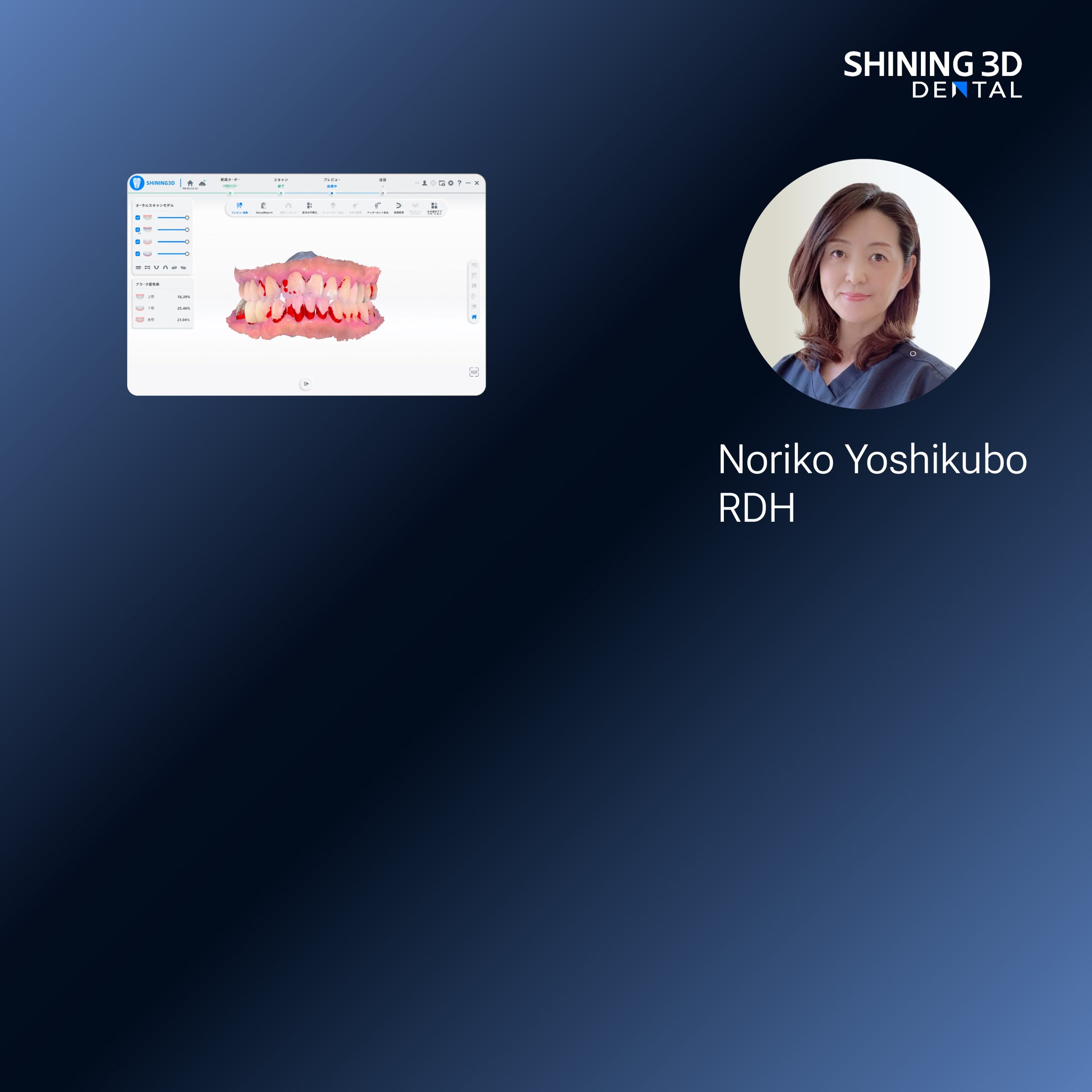
Start with the problem — misfit prosthetics cost time and confidence
Poor-fitting implant restorations frustrate teams and patients: late adjustments, sore soft tissue, repeated appointments. The core fault is often a weak occlusal record or a lost spatial relationship between implants and the patient’s functional jaw position, not the lab’s craftsmanship. Capture a reliable jaw relation record up front and you change the game — dramatically fewer remakes, fewer chairside compromises, and faster confident delivery.
Why photogrammetry solves the repeat offenders
Photogrammetry locks implant positions in three dimensions with surgical-level accuracy. Instead of guessing where the implant analog sat when the patient shut down, you photographically preserve geometry and build the prosthesis to a fixed, measurable coordinate set. That’s the technical edge many clinicians lack when they wrestle with inconsistent implant-to-occlusion transfers.
Practical workflow — clear, actionable steps
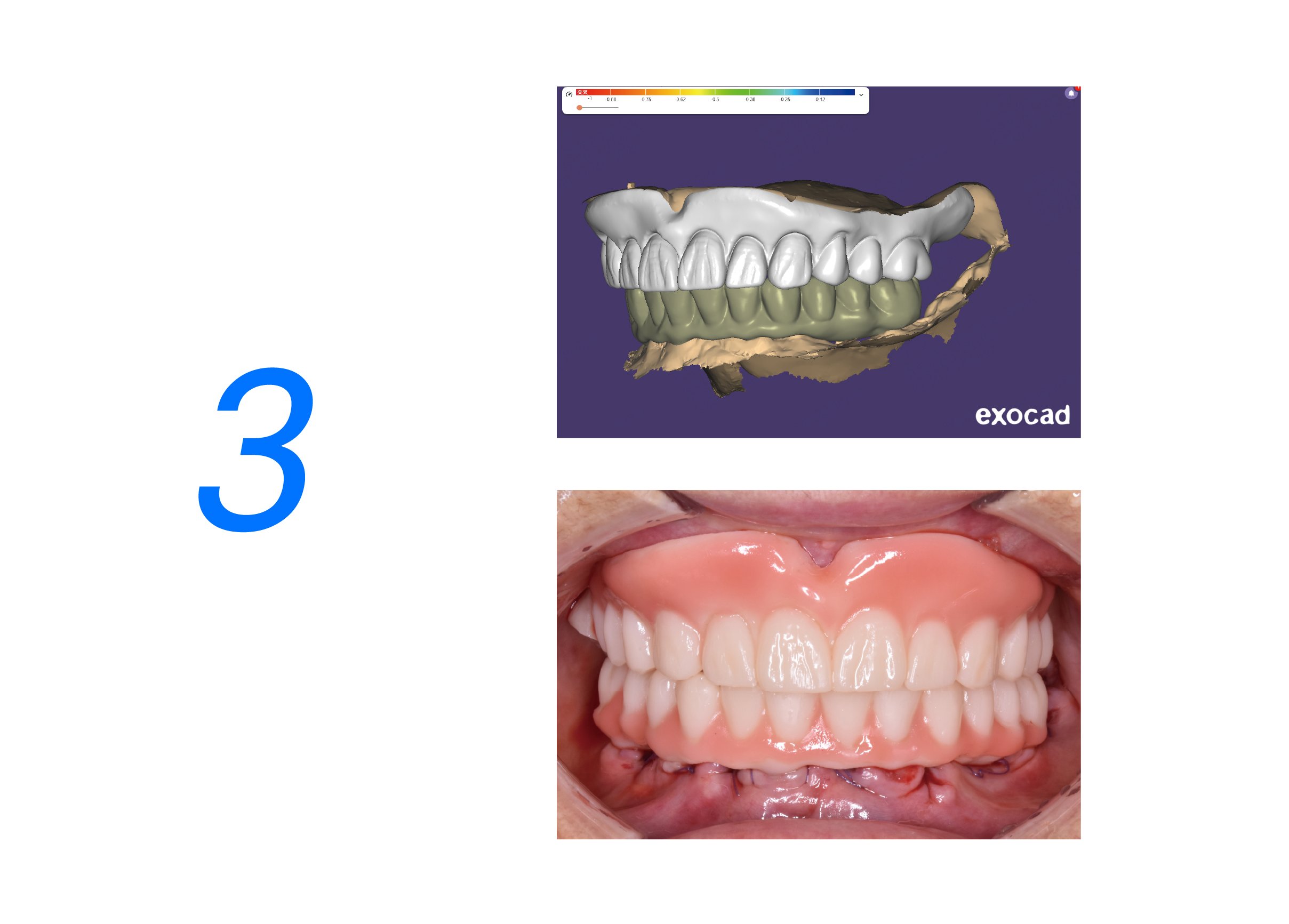
1) Stabilize the patient’s functional jaw position with a validated record. 2) Use photogrammetry to capture implant locations and the opposing arch. 3) Merge the occlusal registration into the implant coordinate system before sending files to the lab. 4) Confirm the virtual assembly with a digital try-in or a printed verification jig. Do these consistently and you remove guesswork from fit and occlusion.
Real experience and credible reference
I’ve worked with restorative teams who cut lab callbacks from multiple visits to just one verification. That outcome aligns with live demonstrations at the International Dental Show (IDS) in Cologne where clinicians showcased integrated jaw motion tracking and photogrammetry setups — practical proof that recording dynamic jaw data alongside static implant geometry improves final function and patient comfort.
Common pitfalls to avoid
Relying on a single wax bite taken after implant placement is risky: tissue bounce and inaccurate seating distort the relation. Don’t skip verification of scanbody seating during photogrammetry capture. Avoid exporting scans without checking the merged occlusal plane. Finally, don’t expect passive implant frameworks to fix active occlusal errors — you must record and design for the functional movements you observe.
Alternatives and when to use them
Traditional analog impressions still have a place for simple, single-tooth cases with predictable occlusion. Full-arch, multi-implant cases benefit most from photogrammetry because of cumulative error in stitched scans. If access, cost, or training prevent immediate adoption, combine high-quality intraoral scanning with meticulous chairside occlusal records as an interim strategy.
Wrap-up — a clear path to fewer remakes and better outcomes
Tackle fit failures by confronting the real source: incomplete spatial and functional records. Use photogrammetry paired with a solid functional record, verify the merged model, and avoid band-aid adjustments. This practical, jaw-aware approach is what leads to consistent first-try fits and confident patients — the exact outcome teams at SHINING 3D DENTAL designed their solutions to support.